
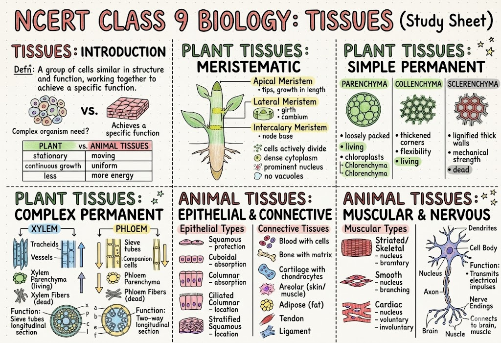
What is a Tissue?
A tissue is a group of cells, similar in structure, that work together to perform a specific function. In multicellular organisms, cells of similar type group together to form a tissue, tissues form organs, organs form organ systems, and organ systems form an organism.
The formation of different types of tissues creates division of labour, which increases efficiency and enables the body to carry out complex life processes. In animals, muscle tissue enables movement and nervous tissue carries messages. In plants, xylem transports water and minerals while phloem transports food.
1. Why are Plant and Animal Tissues Different?
Plants are fixed in one place and need support to stay firm and upright. Their cells have a cell wall that provides rigidity and strength. Animals, in general, can move, and without a rigid cell wall, animal cells can change shape easily — this flexibility suits locomotion.
Another major difference is nutrition. Animals have tissues for digesting food from different sources, while plants have tissues for utilising solar energy through photosynthesis. Growth patterns also vary because tissues responsible for growth differ in structure and function.
2. Tissues for Growth in Plants
Plants grow in three ways:
- Increase in length (height of stem and depth of roots)
- Increase in girth (thickness of stem)
- Regrowth after cutting of branches or grazing by animals
This growth requires actively dividing cells that together form meristematic tissue.
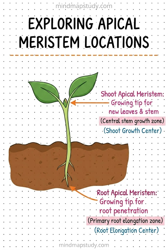
2.1 Apical Meristem — How do Plants Grow in Length?
Plants have growth zones at the tips of their roots and shoots called apical meristems. These help plants grow in length. Root tips contain actively dividing cells (mitosis), and similarly shoot tips contain actively dividing cells that help shoots grow in length.
2.2 Lateral Meristem — How do Plants Grow in Girth?
The stems of dicot plants not only grow in length but also increase in diameter or girth over time. This happens due to actively dividing cells arranged in a ring in the stem. These cells divide and produce new cells inside and outside in a concentric manner, leading to an increase in the diameter of the stem. This meristematic tissue is called the lateral meristem.
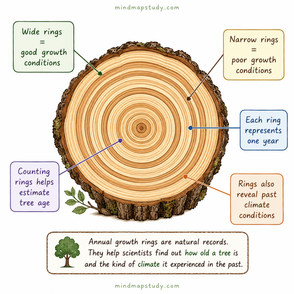
If you observe the cut surface of a tree trunk, you will notice ring-like patterns called annual growth rings. Wide rings indicate favourable growth conditions and narrow rings indicate unfavourable conditions. By counting these rings, scientists can estimate the age of a tree and understand the climatic conditions under which it grew.
2.3 Intercalary Meristem — How do Plants Grow After Being Cut?
When the tip of a young stem is cut, the stem stops growing in length but new branches arise from the nodes of the stem. The intercalary meristem is located at the base of an internode or just above the node.
- The node is the point on a plant stem where branches or leaves arise.
- The internode is the part of stem between two nodes.
Grasses regrow after being mowed or grazed because of intercalary meristem present at the nodes. Similarly, hedges become bushy again after cutting due to this tissue.
Summary of Three Meristematic Tissues:
| Type | Location | Function |
|---|---|---|
| Apical meristem | Root and shoot tips | Increases length |
| Lateral meristem | Along the circumference of stems | Increases girth |
| Intercalary meristem | Base of internode / just above node | Regrowth after cutting |
Imp characteristics of meristematic tissue cells:
- Small in size
- Thin cell walls
- Large and prominent nucleus
- Dense cytoplasm with many organelles
- Vacuoles are generally absent
- Cells are tightly packed with little or no intercellular space
These characteristics allow continuous and rapid cell division.
2.4 Permanent Tissues
Due to continuous cell division, meristematic tissue adds new cells to the plant body. Some newly formed cells remain meristematic while others lose the ability to divide. Cells that lose the ability to divide undergo changes in structure and function and become permanent tissues. They become specialised to perform specific functions such as support, transport, or storage.
The process by which meristematic tissue becomes specialised to perform specific functions is called differentiation.
Permanent tissues can be:
- Simple — composed of only one type of cell
- Complex — composed of more than one type of cell
(i) Protective Tissue — Epidermis
The epidermis forms the outermost layer of the plant body. It consists of a tightly packed, single layer of flat and rectangular cells and protects all parts of the plant.
- Cells are covered with a waxy layer of cutin called cuticle, which protects against mechanical injury, invasion by parasites, and reduces water loss.
- In plants living in very dry habitats, the epidermis may be covered by a thick layer of cuticle to reduce water loss through transpiration.
- In roots, hair-like projections called root hair arise from epidermal cells. These increase surface area for absorption of water and minerals from soil.
- In leaves, the epidermis contains pores called stomata, which help in gaseous exchange and transpiration (evaporation of water vapours). Transpiration creates a transpiration pull in xylem and also helps in elimination of wastes.
(ii) Supporting Tissue — Simple Permanent Tissues
Three types of simple permanent (supporting) tissues are present in plants:
a. Parenchyma
- Consists of living cells with thin walls
- Loosely packed with intercellular spaces
- Mainly stores food
- Also performs photosynthesis in green parts
- In aquatic plants, specialised parenchyma forms air spaces to help them float
b. Collenchyma
- Consists of living cells with unevenly thickened corners due to pectin deposition
- Pectin gives flexibility like rubber
- Provides support and flexibility, allowing stems and tendrils to bend without breaking
c. Sclerenchyma
- Cells have thick walls due to deposition of lignin, making them hard and strong (forms the woody structure)
- Most cells are dead
- Found in stems, leaf veins, and hard coverings of seeds and nuts such as coconut husk and walnut shell
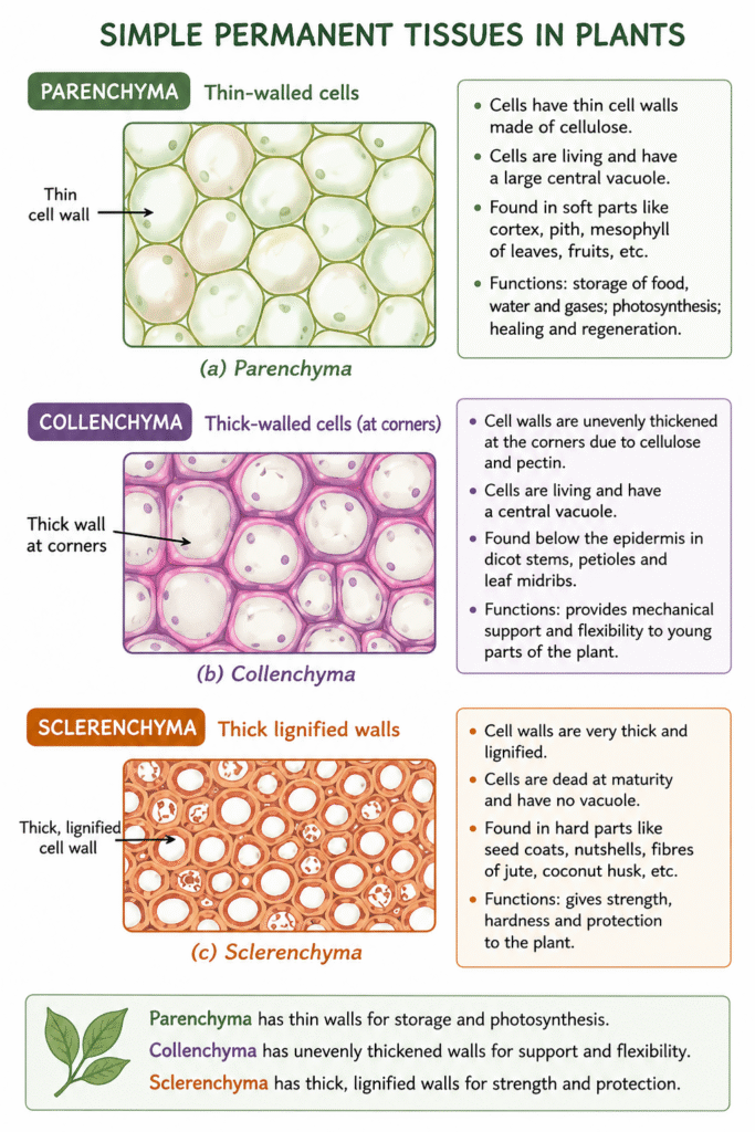
Comparison of Simple Permanent Tissues:
| Feature | Parenchyma | Collenchyma | Sclerenchyma |
|---|---|---|---|
| Cell wall | Thin | Unevenly thickened at corners | Thick, lignified |
| Living/Dead | Living | Living | Mostly dead |
| Intercellular spaces | Present | Few | Absent |
| Main function | Storage, photosynthesis | Flexibility, support | Strength, mechanical support |
(iii) Conducting Tissues — Complex Permanent Tissues
Plants have specialised conducting tissues called xylem and phloem, together called complex permanent tissues because they are made of different types of cells working together.
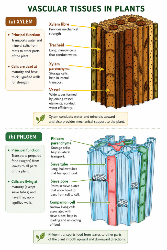
Xylem:
- Transports water and minerals from roots to other parts
- Also provides strength to the plant
- Components: tracheids, vessels, xylem parenchyma, and xylem fibres
- Tracheids and vessels are tubular and thick-walled
- Xylem parenchyma is the only living component; tracheids, vessels, and xylem fibres are primarily sclerenchymatous
Phloem:
- Transports food from leaves to other parts of the plant
- Mostly made up of living cells
- Components: sieve tubes, companion cells, phloem parenchyma, and phloem fibres
- Sieve tubes are long tubular cells joined end to end by perforated walls
- Companion cells regulate cellular functions of sieve tube cells; main function is to monitor loading and unloading of sugars in sieve tubes
- Phloem parenchyma stores food materials, resin, tannins, and latex
- Phloem fibres are primarily sclerenchymatous and provide strength
Plant Tissue Systems
Plant tissues are organised into three tissue systems:
- Dermal tissue system — Forms the outer covering; protects inner parts and reduces water loss
- Ground tissue system — Forms the main body of the plant between dermal and conducting tissues; includes parenchyma, collenchyma, and sclerenchyma
- Vascular tissue system — Consists of conducting tissues — xylem and phloem
3. Animal Tissues
Animal cells also group together and specialise in performing different functions. There are four main types of animal tissues: epithelial, connective, muscular, and nervous.
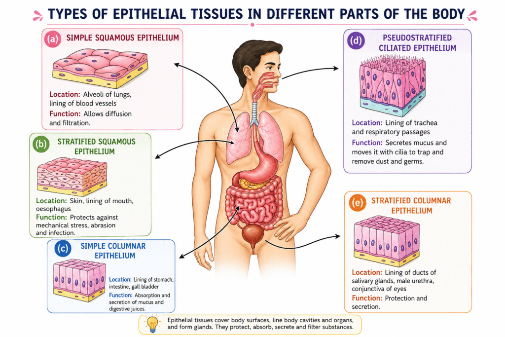
3.1 Epithelial Tissues — Structure and Functions
Epithelial tissue forms the outer covering of the body (skin) and also lines internal organs such as the mouth, lungs, blood vessels, and intestine. It is composed of closely packed cells with very little space between them. This structure prevents entry of germs, reduces water loss, and helps in absorption, secretion, and movement of substances.
Types of Epithelial Tissues:
| Function | Structure | Location in the Body |
|---|---|---|
| Exchange: rapid diffusion of liquids and gases | Single layer of thin, flat cells | Lining of blood vessels and lungs |
| Protection: from mechanical injury, friction, microbes | Many layers; outer cells flat and tightly packed | Skin, mouth, oesophagus |
| Secretion: mucus, enzymes, hormones, sweat, saliva | Cells specialised for producing and releasing substances; cuboidal or columnar | Salivary glands, sweat glands, stomach lining |
| Sensory functions: smell, taste, sound, balance | Specialised receptor cells with hair-like cilia | Nostrils, taste buds, inner ear |
| Absorption: nutrients, water | Single layer of tall, pillar-like cells, often with hair-like structure | Lining of small intestine |
3.2 How are Various Parts Connected in Our Body? — Connective Tissue
A tissue that connects and supports other tissues is called a connective tissue. Both blood and bones are connective tissues, though they differ in composition and consistency. Blood is fluid, while bone is hard. This difference is due to the matrix — watery and jelly-like in blood but hard, solid, and rigid in bones.
Components of Blood:
- Red Blood Cells (RBCs) — contain haemoglobin, an iron-rich protein that gives blood its red colour; live for about 4 months and are replaced regularly
- Platelets — help in blood clotting at the site of injury
- White Blood Cells (WBCs) — collect at infected areas, causing inflammation, redness, swelling, and possible pus formation
- Plasma — makes up 55% of total blood volume; formed elements make up 45%
Other Connective Tissues:
| Connective Tissue | Structure/Matrix | Function |
|---|---|---|
| Bone | Rigid matrix of calcium and phosphorus compounds | Gives strength, support, and protection |
| Cartilage | Soft, jelly-like matrix | Provides flexibility and cushions ends of bones for shock absorption |
| Tendon | Tough connective tissue | Connects muscle to bone; brings about movement |
| Ligament | Tough connective tissue | Connects bone to bone; provides stability, limits movement, prevents dislocation |
3.3 Can We Control Movement in Our Body? — Muscular Tissue
Skeletal (Voluntary) Muscles:
- Movements under conscious control (running, writing, lifting)
- Attached to the skeleton
- Made up of bundles of long, cylindrical cells called muscle fibres
- Cells are unbranched, multinucleate (many nuclei), and striated (showing light and dark bands)
Smooth (Involuntary) Muscles:
- Movements not under conscious control (movement of food in intestine)
- Found in organs like stomach and intestines
- Cells are spindle-shaped, have a single nucleus, and lack striations
- Help in slow, continuous movements like digestion
Cardiac Muscles:
- Found only in the heart
- Fibres are cylindrical and branched with a single nucleus
- Have faint striations
- Work tirelessly and rhythmically; enable the heart to beat throughout life without fatigue
Comparison of Muscle Types:
| Feature | Skeletal Muscle | Smooth Muscle | Cardiac Muscle |
|---|---|---|---|
| Control | Voluntary | Involuntary | Involuntary |
| Shape | Long, cylindrical, unbranched | Spindle-shaped | Cylindrical, branched |
| Nucleus | Multinucleate | Single | Single |
| Striations | Present (light and dark bands) | Absent | Faint striations |
| Location | Attached to skeleton | Stomach, intestines | Heart only |
3.4 How does the Body Sense, Communicate and Respond? — Nervous Tissue
Nervous tissue forms the body’s control and coordination network. The brain acts as the control centre, coordinating activities, memory, and responses. Muscles cannot function independently — they receive instructions from the nervous tissue.
The cells of nervous tissue are called neurons (nerve cells), which are specialised to receive, process, and transmit messages.
Parts of a Neuron:
- Cell body — contains the nucleus and controls cell activities
- Dendrites — receive signals from other neurons
- Axon — a long fibre that carries messages from the cell body and ends at axon terminals
- Axon terminals — transmit messages to other cells
4. The Musculoskeletal System
The musculoskeletal system is made up of bones, muscles, joints, cartilage, tendons, and ligaments. It helps us stand upright, move, maintain posture, and protect delicate organs. It functions under the control of the nervous system.
Muscles pull on bones to produce movement. They are attached to bones by strong, flexible bands called tendons. When a muscle contracts, the tendon transmits this force to the bone, resulting in movement at a joint.
On average, the adult human skeleton makes up about 12–15% of body weight (can vary with age, gender, and body composition).
4.1 The Musculoskeletal System in Action
A joint is a junction between two or more bones. Joints allow movement but cannot move bones on their own — muscles do that.
5. Types of Joints
5.1 Ball and Socket Joint
The rounded top of the upper arm bone fits into a shallow hollow of the shoulder bone. This joint allows forward, backward, sideways, and circular movements. The shoulder forms the shoulder girdle together with the collarbone, connecting the arm to the skeleton.
5.2 Hinge Joint
The elbow bends and straightens in one direction only, like a door hinge. A similar hinge joint is present in the knee, where a small bone called the kneecap protects the joint.
5.3 Pivot Joint
The skull is connected to the backbone through a pivot joint, which allows the head to move side to side.
5.4 Fixed Joints
The bones of the skull are connected by fixed joints — they cannot move. This keeps the brain safe.
Summary of Joint Types:
| Joint Type | Location | Movement Allowed |
|---|---|---|
| Ball and socket | Shoulder, hip | Forward, backward, sideways, circular |
| Hinge | Elbow, knee | Bending in one direction only |
| Pivot | Neck (skull and backbone) | Side-to-side rotation |
| Fixed | Skull | No movement |
6. Skeletal System
The skeletal system consists of a framework of bones that provides strength and protects delicate internal organs. It includes:
- Skull — hard case of flat bones joined by fixed joints to protect the brain, eyes, and ears
- Vertebral column (backbone/spine) — made up of small bones called vertebrae; supports the body and helps us stand upright. Between each vertebra is a cartilage disc that acts as a cushion and allows flexibility so we can bend and twist without injuring the spinal cord.
- Rib cage — 12 pairs of ribs forming a protective cage for the heart and lungs. Ribs are attached to the spine at the back and to the sternum (breast bone) in front, joined by flexible cartilage. This flexibility allows the rib cage to expand and contract during breathing.
Exercise Questions — Solutions
Q1. Meristematic tissues divide repeatedly. What property of their cells allows them to do this?
Answer: (iii) They have thin walls, dense cytoplasm and large prominent nucleus.
Meristematic cells are small with thin cell walls, dense cytoplasm, and a large prominent nucleus. Vacuoles are absent. These structural features allow continuous and rapid cell division.
Q2. If a plant is unable to transport food from leaves to roots, which tissue is malfunctioning?
Answer: (ii) Phloem
Phloem is responsible for transporting food (sugars) prepared in the leaves to all other parts of the plant, including the roots. This is called translocation.
Q3. Why are the epithelial tissues that line an animal’s internal organs usually only one or a few cells thick?
Answer: (iii) To allow quick exchange of materials across them.
A thin epithelial layer allows rapid diffusion of gases (like in lungs) and quick absorption of nutrients (like in the intestine). Multiple layers would slow down this exchange.
Q4. You can perform these two jumps — Straight-leg jump (keep knees and ankles stiff) and Normal jump (bend knees and ankles naturally). How did your ankle, knee, and hip positions differ between the two jumps?
In a straight-leg jump, the knees and ankles remain stiff and do not bend. The hips may bear the full force of landing. The jump is shorter and landing is harder and more jarring because the joints do not absorb the shock.
In a normal jump, the knees bend (flex) during takeoff and especially at landing, the ankles flex downward during the jump and absorb impact on landing, and the hips also bend slightly to lower the centre of gravity. The joints — especially the hinge joints at the knee and ankle — work together to absorb the landing force, making the movement smoother and safer.
Q5. Which type of joint is involved when you bend your knees and ankles?
Answer: (ii) Hinge
The knee and ankle are hinge joints, which allow bending and straightening movement in one direction only, similar to how a door hinge works.
Q6. Assertion-Reason Questions:
A. Assertion: Epithelium is well-suited for gas exchange in the lungs. Reason: It consists of multiple layers of tall cells that slow down diffusion.
Answer: (iii) (A) is true, but (R) is false.
The assertion is correct — epithelium in the lungs is well-suited for gas exchange. However, the reason is false. The epithelium in the lungs consists of a single layer of thin, flat cells (not multiple layers of tall cells), which allows rapid diffusion of gases.
B. Assertion: Cardiac muscle can contract continuously without fatigue. Reason: Cardiac muscle cells have a high number of mitochondria and an abundant blood supply.
Answer: (i) Both (A) and (R) are true, and (R) is the correct explanation of (A).
Cardiac muscle works tirelessly and rhythmically throughout life. This is possible because cardiac muscle cells are richly supplied with mitochondria (producing energy continuously) and receive a constant blood supply delivering oxygen and nutrients.
C. Assertion: Tendons connect bone to bone and allow joint movement. Reason: Tendons are made of tough connective tissue that transmits force from muscle to bone.
Answer: (iv) (A) is false, but (R) is true.
The assertion is false — tendons connect muscle to bone (not bone to bone). Ligaments connect bone to bone. However, the reason is correct — tendons are indeed made of tough connective tissue that transmits the force of muscle contraction to the bone.
D. Assertion: In a hinge joint, movement occurs primarily in one plane. Reason: The bone ends are shaped to allow sliding in all directions.
Answer: (iii) (A) is true, but (R) is false.
The assertion is correct — hinge joints (like the elbow and knee) allow movement in only one plane (bending/straightening). The reason is false — the bone ends in a hinge joint are shaped to allow movement in one direction only, not in all directions.
Q7. Plot a graph between the age of a tree (in years) on the x-axis and the diameter of the tree (in cm) along with the number of annual rings formed over time on the y-axis.
(i) Analyse the graph in terms of the diameter of the stem over time:
The diameter of the teak tree increases steadily with age. Between year 10 and year 20, there is a notably sharper increase (from 8 cm to 24 cm), suggesting a period of faster growth. Overall, the graph shows a positive, increasing relationship between age and stem diameter.
(ii) What is the relation between the diameter of the teak tree and the annual rings formed?
There is a direct relationship between the diameter of the teak tree and the number of annual rings. As the number of annual rings increases, the diameter also increases. In fact, the number of annual rings equals the age of the tree in years, and both correspond to growth in girth over time.
(iii) Which specialised tissue is responsible for the girth of the stem and where is it located?
The lateral meristem is responsible for the increase in girth of the stem. It is located along the circumference of the stem (arranged in a ring). It divides and produces new cells inside and outside in a concentric manner, leading to an increase in diameter. Each year’s growth produces one annual ring.
Q8. A tree was severely debarked by an elephant. Based on your learning, answer the following:
(i) Which functions of the tree are hampered by debarking?
The bark contains cork cells and the outer protective layer (epidermis/cork). Debarking removes this protective covering, so the following functions are hampered:
- Protection from mechanical injury, pathogens, and desiccation is lost
- Regulation of water loss is disrupted
- If phloem lies just beneath the bark, food transport from leaves to roots is also disrupted
(ii) Which plant tissue would be affected by further damage to the tree trunk even after debarking?
Further damage to the trunk would affect the lateral meristem (responsible for secondary growth and girth) and the xylem and phloem (the vascular tissues responsible for transporting water, minerals, and food).
(iii) Which function of the tree would be hampered if the tissues beneath the bark were severely damaged?
If tissues beneath the bark (xylem and phloem) are severely damaged:
- Transport of water and minerals from roots to leaves via xylem would stop
- Transport of food from leaves to roots via phloem would stop
- The tree would not be able to grow or survive
(iv) What assumptions are you making to answer the questions above? How would the answer change if your assumptions are also changed?
Assumptions made:
- The bark includes the cork/epidermis layer and that phloem lies just beneath it
- The tree is a dicot with a well-defined bark, vascular cambium, xylem, and phloem
- Damage is complete in the debarked region (ring-barking)
If assumptions change — for example, if only partial debarking occurs, some protective function would remain. If the phloem is not damaged, food transport would continue. If the tree is a monocot, the arrangement of vascular tissue differs and the impact would be different.
Q9. Aamrapali observed that a young mango sapling’s stem bends flexibly during monsoon winds and does not break. Which tissue is responsible for this flexibility? Predict and provide your explanation of the impact if the existing tissue was replaced by sclerenchyma.
The tissue responsible for the flexibility of the young mango sapling’s stem is collenchyma. Collenchyma cells have unevenly thickened corners due to pectin deposition, which gives them flexibility similar to rubber, allowing the stem to bend without breaking.
If collenchyma were replaced by sclerenchyma, the stem would become very hard and rigid because sclerenchyma cells have thick, lignified walls and are mostly dead. During strong monsoon winds, the stem would no longer be able to bend — it would break instead of bending. The sapling would not survive mechanical stress, and the overall flexibility and resilience of the young plant would be completely lost.
Q10. Sohan designed an experiment for the regeneration of sugarcane using two types of cuttings, type ‘A’ and type ‘B’. Type ‘B’ sprouted while type ‘A’ did not.
(i) Why were type ‘B’ cuttings able to grow as sugarcane but type ‘A’ could not?
Type ‘B’ cuttings contained a node, which houses the intercalary meristem. This meristematic tissue has actively dividing cells capable of regenerating new growth. Type ‘A’ cuttings most likely contained only the internode portion (without a node), and therefore lacked the meristematic tissue needed for sprouting.
(ii) What difference was present in type ‘B’ compared to type ‘A’?
Type ‘B’ cuttings had at least one node present, while type ‘A’ cuttings were only internodal segments without a node. The presence of the node (with intercalary meristem) is what allowed type ‘B’ to sprout and grow.
(iii) What observation or measurement was made to determine whether this change had an effect?
The observation was whether sprouting occurred or not after a few weeks. The presence or absence of new growth (new shoots emerging from the cuttings) was the measurement used to determine the effect.
(iv) What parameters should be kept the same for both types of cuttings to ensure a fair comparison?
- Same species and variety of sugarcane
- Same length and thickness of cuttings
- Same soil type and quantity
- Same amount of water and sunlight
- Same temperature and humidity conditions
- Same time period of observation
- Same number of cuttings used in each group
Q11. Rohan says “A tissue is a group of similar cells performing similar functions.” Rajiv counter argues that this is true for simple tissues but different for complex tissues. Provide your explanation.
Both Rohan and Rajiv are partially correct.
Rohan’s statement holds true for simple permanent tissues like parenchyma, collenchyma, and sclerenchyma, where all cells are of one type and perform the same function — for example, parenchyma cells all store food.
However, Rajiv is right regarding complex permanent tissues like xylem and phloem. These are made up of more than one type of cell working together. For instance:
- Xylem contains tracheids, vessels, xylem parenchyma, and xylem fibres — different cell types with different roles (conduction, strength, storage)
- Phloem contains sieve tubes, companion cells, phloem parenchyma, and phloem fibres — each playing a different role in food transport
So while the definition of tissue as “a group of similar cells” broadly holds, complex tissues show that different cell types can work together as a unit to perform a common overall function. The correct and complete definition should acknowledge that a tissue is a group of cells (similar or different) working together to perform a specific function.
Q12. Coconut husk fibres are used for mats which are tough and fibrous. Which tissue has structural features suitable for providing this strength? Explain why living parenchyma couldn’t serve the same purpose.
The tissue responsible for the toughness and fibrous nature of coconut husk is sclerenchyma. Sclerenchyma cells have thick, lignified walls that make them extremely hard, strong, and rigid. Most sclerenchyma cells are dead at maturity, meaning there is no metabolic activity and all space is occupied by the cell wall, making the fibre strong and durable.
Living parenchyma cells cannot serve the same purpose because:
- Their walls are thin and lack lignin
- They are living cells with large vacuoles and active metabolism
- They are loosely packed with intercellular spaces
- They provide no significant mechanical strength
Parenchyma is suited for storage and photosynthesis, not for providing the structural rigidity needed in products like mats or ropes.
Q13. Vibha claims “Meristematic cells are located only at the root and shoot apices.” Is this correct? What question can Neha ask Vibha to help her understand further?
Vibha’s statement is incorrect. Meristematic cells are present in three locations, not just the root and shoot tips:
- Apical meristem — at root and shoot tips (this is what Vibha mentioned)
- Lateral meristem — along the circumference of the stem (responsible for increase in girth)
- Intercalary meristem — at the base of internodes or just above the nodes (responsible for regrowth after cutting, as seen in grasses)
Neha could ask Vibha: “Have you ever noticed that grass regrows even after being mowed or grazed by animals — if meristematic cells are only at the tips, which meristem is responsible for this regrowth, and where exactly is it located?”
This question would guide Vibha to think about intercalary and lateral meristems as well.
Q14. A plant cell and an animal cell are of the same size.
(i) Which cell will have a larger vacuole? Give reasons.
The plant cell will have a larger vacuole. Plant cells typically have a large central vacuole that occupies up to 80–90% of the cell’s volume. Vacuoles in plant cells are important for storing water, nutrients, and waste products, and they also help maintain turgor pressure, which keeps the plant firm and upright.
Animal cells, in contrast, either have very small vacuoles or may lack prominent vacuoles altogether. Meristematic plant cells also lack vacuoles, but mature plant cells have large central vacuoles.
(ii) What assumptions are you making to answer the question above?
- Both cells are mature (not meristematic, since meristematic plant cells lack vacuoles)
- The plant cell is a typical mature parenchyma or leaf cell
- The animal cell is a typical body cell (not a specialised cell like a red blood cell, which has no nucleus or vacuole)
- Both cells are from multicellular organisms
Q15. A textbook states “Each plant tissue performs only one specific function.” What questions would you ask to critically examine the correctness of this statement? What examples of tissues would you take?
This statement is an oversimplification and needs critical examination. Questions you could ask:
- Does parenchyma perform only one function, or does it also perform photosynthesis, storage, and even provide buoyancy in aquatic plants?
- Does xylem only transport water, or does it also provide mechanical strength to the plant?
- Do stomata in the epidermis only perform one function, or do they assist in both gaseous exchange and transpiration?
- Can sclerenchyma be considered to perform only one function, or does it serve both support and protection in different plant parts?
Examples of tissues that perform more than one function:
- Parenchyma — stores food, performs photosynthesis in green parts, and in aquatic plants forms aerenchyma for buoyancy
- Xylem — transports water and minerals, and also provides mechanical support to the plant body
- Epidermis — protects the plant, reduces water loss, absorbs water through root hair, and allows gaseous exchange through stomata
These examples show that the textbook statement is not entirely accurate. Most plant tissues perform more than one function, and the statement should be revised to reflect that tissues are primarily specialised for one function but may also carry out additional roles.