
🫀 Life Processes
All concepts explained simply — nutrition, respiration, transport, excretion — with every question answered.
Table of Contents
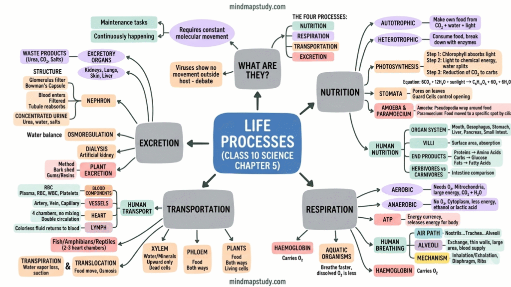
Toggle1 What Are Life Processes?
All living things need to constantly maintain and repair their internal structures. These maintenance tasks — carried out even when we are asleep — are called life processes.
Because all living structures are made of molecules, life requires continuous molecular movement. This is why viruses, which show no molecular movement outside a host cell, are controversial — are they truly alive?
Fig. 1 – The four essential life processes; arrows show how each feeds into the central maintenance of life
Life processes are Nutrition → Respiration → Transportation → Excretion. Energy is needed for all of them. This energy comes from food (outside the body).
2 Nutrition — Autotrophic vs Heterotrophic
Nutrition is the process by which organisms obtain energy and raw materials from outside their bodies for growth, maintenance, and repair.
🌱 Autotrophs
Make their own food using simple inorganic substances (CO₂ and water) and sunlight energy. Examples: green plants and some bacteria. Store energy as starch (plants) or glycogen (animals).
🦁 Heterotrophs
Cannot make their own food. They consume complex substances prepared by other organisms and break them down using enzymes. Examples: animals, fungi. Depend directly or indirectly on autotrophs.
Some heterotrophs break food down outside their body and then absorb it — like fungi (bread mould, mushroom, yeast). Others are parasites that get nutrition from a living host without killing it — like cuscuta (amar-bel), leeches, tapeworms, ticks.
3 Photosynthesis
Photosynthesis is the process by which green plants convert carbon dioxide and water into glucose using sunlight energy and chlorophyll. Unused glucose is stored as starch.
(Carbon dioxide + Water → Glucose + Oxygen + Water)
Three Steps of Photosynthesis
- 1Absorption of light by chlorophyll (green pigment in chloroplasts)
- 2Conversion of light energy to chemical energy and splitting of water molecules into hydrogen and oxygen
- 3Reduction of CO₂ to carbohydrates using the hydrogen and chemical energy
Fig. 2 – Photosynthesis: CO₂ and H₂O enter the leaf (blue arrows ↓); sunlight energy absorbed by chloroplasts; O₂ exits and glucose is stored (green arrows →)
Stomata — How CO₂ Enters
Stomata are tiny pores on the surface of leaves through which CO₂ enters and O₂ exits. Each stoma is surrounded by a pair of guard cells that control its opening and closing.
Guard cells swell (absorb water) → stomatal pore opens. Guard cells shrink (lose water) → pore closes. Plants close stomata when they don't need CO₂ (no photosynthesis) to prevent water loss.
Other Raw Materials for Autotrophs
Besides CO₂ and water, plants also need nitrogen, phosphorus, iron, magnesium and other minerals from the soil. Nitrogen is essential for making proteins. It is absorbed as inorganic nitrates/nitrites or through organic compounds prepared by soil bacteria.
4 Heterotrophic Nutrition — Amoeba
Amoeba is a unicellular organism that feeds using temporary finger-like extensions called pseudopodia. These wrap around a food particle to form a food vacuole. Inside the vacuole, enzymes digest the food into simpler molecules which diffuse into the cytoplasm. Undigested material is expelled out.
Paramoecium is also unicellular but has a fixed shape. Food is moved to a specific spot by cilia (hair-like structures covering its surface).
Fig. 3 – Amoeba feeding: ① detects food → ② pseudopodia engulf it → ③ food vacuole + enzyme digestion → ④ waste egested outward (red arrow)
5 Nutrition in Human Beings
The human digestive system is essentially a long tube called the alimentary canal, running from the mouth to the anus. Different regions are specialised for different functions.
Fig. 4 – Human Alimentary Canal: food travels downward (grey arrows ↓); green/yellow dashed arrows show secretions entering from accessory organs; blue arrow shows absorption into blood; red arrow = waste expelled
Step-by-Step Digestion
| Organ | Secretion | Action |
|---|---|---|
| Mouth | Saliva (salivary amylase) | Amylase breaks starch → simple sugar. Teeth crush food. Tongue mixes. |
| Oesophagus | Mucus | Peristalsis moves food to stomach. No digestion. |
| Stomach | HCl + Pepsin + Mucus | HCl makes acidic medium. Pepsin digests proteins. Mucus protects stomach lining. |
| Liver | Bile juice | Makes food alkaline. Emulsifies fats (breaks into small globules). |
| Pancreas | Pancreatic juice (Trypsin + Lipase) | Trypsin digests proteins. Lipase digests emulsified fats. |
| Small intestine | Intestinal juice | Completes digestion: proteins → amino acids, carbs → glucose, fats → fatty acids + glycerol. Villi absorb nutrients. |
| Large intestine | — | Absorbs water from undigested material. |
| Anus | — | Undigested waste is removed by anal sphincter. |
Villi are finger-like projections lining the small intestine. They massively increase the surface area for absorption. They are richly supplied with blood vessels to carry absorbed nutrients to all body cells.
Grass contains cellulose, which is very tough to digest and needs a longer intestine. Meat (protein) is much easier to digest — so carnivores like tigers have shorter small intestines.
6 Respiration
Respiration is the process of breaking down glucose (food) to release energy inside cells. The energy is stored in molecules of ATP (Adenosine Triphosphate) — the energy currency of the cell.
Fig. 5 – Glucose → Pyruvate (always); then 3 paths: ① Yeast anaerobic (red ←), ② Muscle anaerobic (yellow ↙), ③ Aerobic in mitochondria (green →) — aerobic gives most ATP
| Feature | Aerobic | Anaerobic |
|---|---|---|
| Oxygen needed? | ✅ Yes | ❌ No |
| Location | Mitochondria | Cytoplasm |
| End products | CO₂ + H₂O + Energy | Ethanol+CO₂ (yeast) / Lactic acid (muscles) |
| Energy released | Much more (36–38 ATP) | Very little (2 ATP) |
| Examples | Most animals, plants | Yeast, muscle cells during intense exercise |
ATP (Adenosine Triphosphate) is the energy currency of cells. Respiration releases energy which is used to form ATP from ADP + phosphate. ATP then powers all other cellular activities — muscle contraction, protein synthesis, nerve impulse conduction. Think of ATP like a rechargeable battery.
7 Breathing in Human Beings
Air enters through the nostrils → filtered by fine hairs and mucus → passes through throat → trachea (supported by rings of cartilage) → bronchi → bronchioles → alveoli.
Fig. 6 – Human Respiratory System: blue arrow ↓ = inhaled air path; O₂ crosses into blood (green →); CO₂ exits blood into alveoli (red ←); diaphragm controls breathing
Alveoli — Where Gas Exchange Happens
Alveoli (singular: alveolus) are tiny balloon-like sacs at the end of bronchioles in the lungs. Their walls are one-cell thick and surrounded by a dense network of blood capillaries. The enormous number of alveoli creates a very large surface area (~80 m²!) for gas exchange.
Mechanism: Diaphragm flattens + ribs lift → chest cavity expands → air rushes in (inhalation). O₂ from air crosses alveolar walls into blood. CO₂ from blood crosses into alveoli → exhaled.
Haemoglobin is the red pigment in red blood cells (RBCs) that carries oxygen. It has a very high affinity for O₂. CO₂ is more soluble in water, so it is transported mostly dissolved in blood plasma. Aquatic organisms breathe much faster than land animals because dissolved O₂ in water is much less than O₂ in air.
8 Transportation in Human Beings
The circulatory system transports food, oxygen, hormones, and waste products throughout the body. It consists of: Heart (pump), Blood (fluid), and Blood vessels (tubes).
Blood — What It Carries
🔴 Plasma
Liquid part (~55%). Yellowish. Transports food, CO₂, nitrogenous wastes in dissolved form. Also carries hormones, salts, proteins.
🔵 Red Blood Cells (RBCs)
Contain haemoglobin. Carry oxygen from lungs to all body cells. Largest population of blood cells.
⚪ White Blood Cells (WBCs)
Fight infections and diseases. Part of the immune system.
🟡 Platelets
Tiny cells that clot the blood at points of injury to prevent excessive bleeding and maintain pressure in blood vessels.
Blood Vessels — Arteries vs Veins
| Feature | Artery | Vein | Capillary |
|---|---|---|---|
| Direction | Heart → Body | Body → Heart | Artery → Vein |
| Pressure | High | Low | Very low |
| Wall thickness | Thick, elastic | Thin | One-cell thick |
| Valves? | No | Yes (prevent backflow) | No |
| Function | Carry oxygenated blood | Carry deoxygenated blood | Exchange materials with cells |
Lymph (Tissue Fluid)
Lymph is a colourless fluid similar to plasma but with less protein. It forms when some plasma leaks out of capillary walls into spaces between cells. Lymph drains into lymphatic capillaries → lymph vessels → back into veins. Lymph carries digested fat from the intestine and drains excess fluid from tissues.
Blood Pressure
Blood pressure is the force blood exerts on the walls of blood vessels. It is measured with a sphygmomanometer.
Normal: 120/80 mm Hg (Systolic/Diastolic). High blood pressure = hypertension (caused by narrow arterioles). Can lead to artery rupture and internal bleeding.
9 The Heart & Double Circulation
Fig. 7 – Double Circulation: Blue arrows = deoxygenated blood path (body → right heart → lungs); Red arrows = oxygenated blood path (lungs → left heart → body)
How Blood Flows Through the Heart
- 1Deoxygenated blood from body arrives at Right Atrium via vena cava → Right Atrium contracts → blood moves to Right Ventricle
- 2Right Ventricle pumps deoxygenated blood to lungs via pulmonary artery for oxygenation
- 3Oxygenated blood from lungs arrives at Left Atrium via pulmonary veins → Left Atrium contracts → blood moves to Left Ventricle
- 4Left Ventricle pumps oxygenated blood to the whole body via aorta
In humans (and birds, mammals), blood passes through the heart twice in one complete cycle — once for lungs (pulmonary) and once for body (systemic). This ensures oxygenated and deoxygenated blood never mix, giving maximum efficiency. Fish have only 2 chambers — blood passes through heart only once (single circulation). Amphibians and reptiles have 3 chambers — some mixing occurs.
10 Transportation in Plants
Plants have two separate conducting tubes in their vascular tissue for transport: Xylem (water + minerals) and Phloem (food/photosynthates).
Fig. 8 – Xylem (blue ↑): water + minerals move upward only from roots to leaves via root pressure and transpiration pull. Phloem (green ↕): food moves in both directions from leaves to all parts using ATP energy
Water Transport — How Water Rises in Tall Trees
Step 1 — Root pressure: Roots actively absorb ions from soil → water enters by osmosis → creates an upward push in xylem.
Step 2 — Transpiration pull (main force during day): Water evaporates from leaf cells through stomata. This creates suction that pulls water up from the xylem vessels all the way from the roots. Transpiration = loss of water from aerial parts as vapour.
Food Transport (Translocation)
Translocation is the transport of soluble photosynthesis products (mainly sucrose) from leaves to other parts of the plant through phloem. Sucrose is loaded into phloem using ATP energy, which increases osmotic pressure, pushing food in whichever direction the plant needs — towards roots (spring, for growth) or towards fruits (reproduction).
11 Excretion in Human Beings
Excretion is the removal of harmful metabolic waste products from the body. The main nitrogenous waste in humans is urea (formed in the liver from breakdown of proteins). The excretory system includes: 2 kidneys, 2 ureters, 1 urinary bladder, 1 urethra.
Fig. 9 – Nephron: Red arrow = blood IN via renal artery; blood filtered in glomerulus; filtrate travels through tubule; green arrows = reabsorption back to blood; grey arrow ↓ = urine to collecting duct → ureter; blue arrow = clean blood out
Each kidney has millions of nephrons. The initial filtrate formed daily is about 180 litres, but only 1–2 litres of urine is actually excreted — the rest is reabsorbed. The amount of water in urine depends on body's water level and how much waste needs to be dissolved.
If kidneys fail, an artificial kidney (dialyser) filters blood using a semi-permeable membrane and dialysis fluid. Waste products diffuse out of the blood. Unlike real kidneys, there is no reabsorption in dialysis. This process is called haemodialysis.
12 Excretion in Plants
Plants use very different strategies for waste removal compared to animals. They do not need specialised excretory organs.
🌬️ Gas Exchange
O₂ (waste from photosynthesis) and CO₂ (waste from respiration) are released through stomata and lenticels. Excess water is removed as vapour by transpiration.
🍂 Leaf Fall
Waste products are stored in cell vacuoles or in leaves. When leaves fall off, these stored wastes are removed from the plant.
🌳 Resins & Gums
Some wastes are stored as resins and gums, especially in old xylem tissue. Trees like pine produce these naturally.
🌱 Into Soil
Plants also excrete some waste substances into the soil around their roots.
13 All Questions & Answers
• Organisms make their own food from inorganic materials (CO₂ + H₂O) using sunlight energy and chlorophyll.
• Food is synthesised from simple molecules.
• Examples: Green plants, algae, some bacteria.
Heterotrophic Nutrition:
• Organisms cannot make their own food and depend on other organisms.
• Complex food is obtained from outside and broken down by enzymes.
• Examples: Animals, fungi, most bacteria, parasites.
• Carbon dioxide (CO₂): From the surrounding air, entering through stomata on leaves.
• Water (H₂O): Absorbed from the soil through roots via root hair cells; transported up through xylem.
• Sunlight (energy): From the sun, absorbed by chlorophyll in chloroplasts.
• Chlorophyll: Present in chloroplasts inside green leaf cells (plant makes it itself).
• Minerals (N, P, Fe, Mg etc.): Absorbed from soil through roots.
(1) Creates an acidic medium (low pH) necessary for the enzyme pepsin to work effectively — pepsin can only digest proteins in an acidic environment.
(2) Kills harmful bacteria present in the food we eat, preventing infections.
(Note: Mucus protects the stomach lining from damage by HCl. When too much acid is produced → acidity.)
• Salivary amylase (mouth): Starch → simple sugars
• Pepsin (stomach): Proteins → smaller peptides
• Trypsin (pancreas): Proteins → amino acids
• Lipase (pancreas): Emulsified fats → fatty acids + glycerol
• Intestinal enzymes: Final conversion — proteins → amino acids, carbs → glucose, fats → fatty acids + glycerol
Without enzymes, food cannot be absorbed through the intestinal walls.
(1) Length: Very long (6–7 m), coiled into a compact space — more time for absorption.
(2) Villi: Numerous finger-like projections (villi) lining the inner surface — greatly increase the surface area for absorption.
(3) Rich blood supply: Each villus is richly supplied with blood capillaries — absorbed nutrients immediately enter the bloodstream and are carried to all body cells.
(4) Lacteals: Lymph vessels (lacteals) in each villus absorb fatty acids and glycerol.
• Terrestrial organisms can absorb oxygen more efficiently from air.
• They breathe much slower than aquatic organisms.
• They do not need to work as hard to obtain oxygen.
Aquatic organisms like fish must continuously pump large volumes of water over their gills to extract enough dissolved oxygen, which requires more energy and effort.
(1) Aerobic respiration (in mitochondria, with O₂):
Pyruvate → CO₂ + H₂O + large amount of energy (38 ATP). Example: most animals and plants.
(2) Anaerobic respiration — Fermentation (in yeast, no O₂):
Pyruvate → Ethanol + CO₂ + small energy (2 ATP).
(3) Anaerobic respiration — Muscle cells (during intense exercise, lack of O₂):
Pyruvate → Lactic acid + small energy. Lactic acid build-up causes muscle cramps.
Oxygen transport: O₂ from alveoli binds with haemoglobin in red blood cells to form oxyhaemoglobin. Blood carries it to all body cells, where O₂ is released.
Carbon dioxide transport: CO₂ is more soluble in water, so it is mostly transported in the dissolved form in blood plasma. A small amount is also carried by haemoglobin. CO₂ is released in the alveoli and exhaled.
(1) Branching structure: Airways divide repeatedly — trachea → bronchi → bronchioles — reaching all parts of the lung.
(2) Alveoli: Millions of tiny balloon-like sacs (alveoli) at the end of bronchioles. The total alveolar surface area is about 80 m² — enormous compared to body surface area.
(3) Thin walls: Alveolar walls are just one cell thick for rapid gas diffusion.
(4) Rich blood supply: Dense network of capillaries surrounds each alveolus for efficient gas exchange.
(5) Residual air: Lungs always contain some air even after exhalation — gives continuous time for O₂ absorption.
(1) Heart: A muscular pumping organ that pushes blood throughout the body. It has 4 chambers — 2 atria (collecting) and 2 ventricles (pumping).
(2) Blood: Fluid that actually transports substances. Contains: Plasma (carries dissolved food, CO₂, wastes), RBCs (carry O₂ via haemoglobin), WBCs (fight infection), Platelets (clotting).
(3) Blood Vessels: Network of tubes — Arteries (carry blood away from heart, thick elastic walls), Veins (carry blood to heart, have valves), Capillaries (one-cell thick, site of exchange with tissues).
(4) Lymphatic system: Lymph vessels carry lymph (tissue fluid with fats) back to bloodstream.
If oxygenated and deoxygenated blood mix (as in 3-chambered hearts of amphibians/reptiles), the blood sent to the body would have less oxygen. Separating them in a 4-chambered heart ensures pure oxygenated blood always reaches the body and pure deoxygenated blood always goes to the lungs. This gives maximum efficiency — essential for high-energy animals.
(1) Xylem: Transports water and dissolved minerals from roots upward to leaves and other parts. Made of vessels and tracheids (dead cells). Driven by root pressure and transpiration pull.
(2) Phloem: Transports dissolved food (products of photosynthesis — mainly sucrose) from leaves to all other parts of the plant in both upward and downward directions. Made of living sieve tubes with companion cells. Uses ATP energy (active transport).
(1) Root pressure: Roots actively absorb mineral ions → water enters by osmosis → creates pressure that pushes water upward into xylem. More important at night.
(2) Transpiration pull (main force during day): Water evaporates from leaf cells through stomata → this suction pulls water up the xylem columns from the root. The continuous column of water from root to leaf is maintained by the cohesion (sticking together) of water molecules.
(1) Sucrose is actively loaded into phloem sieve tubes using ATP energy.
(2) This increases osmotic pressure in phloem → water enters phloem from xylem by osmosis.
(3) The increased pressure drives food through phloem to areas of lower pressure (wherever the plant needs it — roots, fruits, seeds, growing buds).
(4) Transport occurs in both directions (up and down) in sieve tubes with help of companion cells.
• Glomerulus: A knot of capillaries where blood is filtered under pressure.
• Bowman's capsule: Cup-shaped structure surrounding the glomerulus that collects the filtrate.
• Renal tubule: Long coiled tube where selective reabsorption occurs. Blood capillaries surround this tube.
• Collecting duct: Collects urine from many nephrons and leads to the ureter.
Functioning (3 steps):
Step 1 — Filtration: Blood is filtered in the glomerulus. All small molecules (water, glucose, urea, amino acids, salts) pass into Bowman's capsule. Blood cells and large proteins stay in blood.
Step 2 — Reabsorption: As filtrate flows through the tubule, useful substances (glucose, amino acids, most water, salts) are selectively reabsorbed back into blood capillaries.
Step 3 — Urine formation: What remains (urea, excess water, excess salts) = urine. It flows into collecting duct → ureter → bladder → urethra.
(1) Gaseous wastes: O₂ (waste of photosynthesis) and CO₂ (waste of respiration) exit through stomata and lenticels.
(2) Water: Excess water released as vapour through transpiration via stomata.
(3) Cell vacuoles: Many waste products are stored in cell vacuoles harmlessly.
(4) Leaf fall: Wastes accumulated in leaves are removed when leaves fall off seasonally.
(5) Resins and gums: Some wastes stored as resins and gums in old xylem (e.g., pine resin).
(6) Into soil: Some waste substances are directly excreted into surrounding soil through roots.
(1) Amount of water in the body: If water intake is high, excess water needs to be removed → more urine (dilute). If water intake is low (dehydrated), less water excreted → less urine (concentrated).
(2) Amount of dissolved waste to excrete: More urea/waste → more water needed to dissolve it → more urine.
(3) Hormonal control: ADH (antidiuretic hormone) from the pituitary gland signals kidneys to reabsorb more water when the body is dehydrated → less urine. When well-hydrated, ADH level drops → less reabsorption → more urine.
The kidneys constantly adjust the amount of water reabsorbed in tubules to maintain the body's water balance.
Kidneys filter nitrogenous waste (urea) from blood and produce urine. They are the primary organs of the excretory system.
Xylem transports water and dissolved minerals from roots upward to all parts of the plant. Food is transported by phloem (not xylem).
Photosynthesis (autotrophic nutrition) requires all three: CO₂ and water (raw materials), chlorophyll (pigment to absorb light), and sunlight (energy source).
Aerobic breakdown of pyruvate (using O₂) occurs in the mitochondria — hence called the "powerhouse of the cell." The initial step (glucose → pyruvate) happens in the cytoplasm.
Step 1 — Emulsification (by bile juice from liver): Bile salts break large fat globules into smaller droplets (emulsification), like soap breaks oil — this increases the surface area of fat available for enzyme action.
Step 2 — Enzymatic digestion (by lipase from pancreas): Pancreatic lipase acts on the emulsified fat droplets and breaks them down into fatty acids and glycerol, which are small enough to be absorbed through the walls of the small intestine.
Fatty acids and glycerol are absorbed by lacteals (lymph capillaries) in the villi and transported via the lymphatic system.
(1) Moistens food: Makes food wet and slippery for easy swallowing and smooth movement through the oesophagus.
(2) Begins starch digestion: Saliva contains the enzyme salivary amylase (ptyalin) which starts breaking down starch into simpler sugars (maltose) — this is the first step of chemical digestion.
(3) Helps in chewing: Softens food and helps the tongue move it around while the teeth crush it.
(4) Cleanses mouth: Helps wash away food particles from teeth, reducing bacterial activity.
(1) Carbon dioxide — enters through stomata from air
(2) Water — absorbed from soil by roots
(3) Sunlight — energy source (photosynthesis happens only in light)
(4) Chlorophyll — pigment to absorb light energy (in chloroplasts of green cells)
By-products of photosynthesis:
(1) Oxygen (O₂) — released as a gas through stomata (by-product of water splitting in light reaction)
(2) Water — small amount of water is also a by-product
Anaerobic Respiration: No O₂ required. Occurs in cytoplasm. Glucose incompletely broken down to ethanol+CO₂ (yeast) or lactic acid (muscle cells). Releases very little energy (2 ATP). Incomplete oxidation.
Organisms using anaerobic respiration: Yeast (fermentation — used in bread making, wine), some bacteria (e.g., Clostridium), parasite worms (tapeworm), human muscle cells during vigorous/sudden exercise.
(1) Enormous number: Millions of alveoli in each lung → huge total surface area (~80 m²).
(2) Very thin walls: Alveolar walls are just one cell thick → gases diffuse rapidly across them.
(3) Moist lining: Moisture dissolves gases, speeding up diffusion.
(4) Dense capillary network: Each alveolus is surrounded by a rich network of blood capillaries for fast exchange.
(5) Balloon-like structure: Expandable, allowing large volumes of air to fill them during inhalation.
(1) Less oxygen delivery to cells — cells cannot perform aerobic respiration efficiently.
(2) Fatigue and weakness — due to reduced energy production.
(3) Breathlessness — body tries to compensate by breathing faster.
(4) Pale skin and dizziness — insufficient blood oxygenation.
(5) Reduced immunity — cells cannot function at their peak.
(6) In severe cases, organ damage due to chronic oxygen shortage.
Circuit 1 — Pulmonary circulation:
Right ventricle → Pulmonary artery → Lungs (picks up O₂, drops CO₂) → Pulmonary vein → Left atrium
Circuit 2 — Systemic circulation:
Left ventricle → Aorta → All body organs (drops O₂, picks up CO₂ and wastes) → Vena cava → Right atrium
Why is it necessary?
(1) Keeps oxygenated and deoxygenated blood completely separated — no mixing.
(2) Ensures fully oxygenated blood reaches all body tissues — vital for high-energy mammals and birds.
(3) Allows different pressures — high pressure for systemic, lower for pulmonary, preventing lung damage.
Phloem: Transports soluble food (sugars, amino acids). Direction: both upward and downward. Driving force: Osmotic pressure created by active loading of sucrose using ATP. Requires energy (ATP). Made of living cells (sieve tubes with companion cells).
Alveoli (Gas exchange):
• Structure: Balloon-like air sacs; very thin (one-cell) walls; surrounded by capillaries.
• Function: O₂ diffuses from air into blood; CO₂ diffuses from blood into air. Exchange is by simple diffusion.
Nephrons (Blood filtration):
• Structure: Glomerulus (capillary knot) + Bowman's capsule + renal tubule; thin capillary walls.
• Function: Blood filtered under pressure → filtrate formed → useful substances (glucose, water) reabsorbed → urine (urea, excess water) formed. Involves filtration AND selective reabsorption.
Download Free Mind Map PDF
This mind map contains all important topics of this chapter
Visit our Class 10 Science page for free mind maps of all Chapters